Digestive System
Article objectives
Suppose you are studying and having trouble concentrating. You decide to eat an apple for energy. How does energy stored in the apple get into your cells? What organs and processes break down the apple into nutrients that the body can use for fuel? What organs and processes let the nutrients enter your bloodstream so they can travel to the cells where they are needed? The basic processes involved are digestion and absorption. The organs involved are the organs of the digestive system.
Organs and Functions of the Digestive System
Organs that make up the digestive system are shown in Figure 1. Most of the organs form the gastrointestinal tract. Other digestive organs are called accessory organs. As you read about the organs below, refer to Figure 1 for reference.
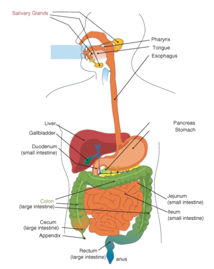
Figure 1: Organs of the digestive system.
Gastrointestinal Tract
The gastrointestinal (GI) tract is a long tube that connects the mouth with the anus. It is more than 9 meters long in adults. The GI tract can be divided into an upper and lower part. The upper GI tract includes the mouth, esophagus, and stomach. The lower GI tract includes the small and large intestines. Food enters the mouth, passes through the upper and lower GI tracts, and then exits the body as feces through the anus.
The organs of the GI tract are covered by two layers of muscles that enable peristalsis. Peristalsis is a rapid, involuntary, wave-like contraction of muscles. It pushes food through the GI tract. The inside of GI tract is lined with mucous membranes. Mucous membranes are moist tissues that can secrete and absorb substances. The ability to secrete and absorb substances is necessary for the functions of the digestive system.
Accessory Organs of the Digestive System
In the lower GI tract, additional organs play important roles in digestion. They are called accessory organs. Food does not pass through them, but they make or store substances needed for digestion. The accessory organs are the liver, gall bladder, and pancreas.
• The liver is a large organ next to the stomach. It produces digestive substances that are carried by ducts, or tubes, to the small intestine and gall bladder.
• The gall bladder is a small, pear-shaped structure below the liver. It stores substances from the liver until they are needed by the small intestine.
• The pancreas is a gland below the stomach. It produces digestive substances that are carried by a duct to the small intestine.
The Liver
The liver is a vital organ that has many functions, including detoxification of blood, protein synthesis, and production of biochemicals necessary for digestion. The liver is also involved in glucose balance. The liver produces bile which breaks down lipids.
The liver performs several roles in carbohydrate metabolism, which help in the balance of blood glucose levels:
• Gluconeogenesis: the synthesis of glucose from certain amino acids, lactate or glycerol • Glycogenolysis: the breakdown of glycogen into glucose • Glycogenesis: the formation of glycogen from glucose.
The liver is one of the most important organs in the body when it comes to blood filtering and detoxification. The liver is involved in getting rid of foreign substances and toxins, especially from the gut. The toxins are usually excreted in bile or urine. Breaking down toxins is referred to as drug metabolism, and is usually done using specialized enzymes produced in the liver. Most of the blood being filtered by the liver is from the portal vein, which carries blood from the intestines. The liver can remove a broad range of microorganisms such as bacteria, fungi, viruses and parasites from the blood. Infections and parasites can come from contaminated water and food, and then find their way into your gut and blood stream. Luckily the blood then goes to the liver for filtering.
The liver also performs several roles in lipid metabolism including cholesterol synthesis and the production of triglycerides (fats). The liver produces coagulation factors I (fibrinogen), II (prothrombin), V, VII, IX, X and XI, as well as protein C, protein S and antithrombin.
Functions of the Digestive System
The digestive system has three main functions: digestion of food, absorption of nutrients, and elimination of solid waste. Digestion is the process of breaking down food into components the body can absorb. There are two types of digestion: mechanical and chemical.
• Mechanical digestion is the physical breakdown of chunks of food into smaller pieces. It takes place mainly in the mouth and stomach.
• Chemical digestion is the chemical breakdown of large, complex food molecules into smaller, simpler nutrient molecules that can be absorbed by the blood. It takes place mainly in the small intestine.
Chemical digestion could not take place without the help of digestive enzymes. Enzymes are substances that speed up chemical reactions. Digestive enzymes speed up the reactions of chemical digestion. Digestive enzymes are secreted by glands in the mucous membranes of the mouth, stomach, small intestine, and pancreas. Different digestive enzymes help break down different types of food molecules, including carbohydrates, proteins, and lipids.
The name of a digestive enzyme typically ends with the suffix -ase, which means “enzyme”. The rest of the name refers to the type of food molecules the enzyme helps digest. For example, the enzyme lipase helps digest lipid molecules, and the enzyme lactase helps digest molecules of the sugar lactose.
After food is digested, the resulting nutrients are absorbed. Absorption is the process in which substances pass into the blood stream, where they can circulate throughout the body. Absorption occurs mainly in the small intestine. Any remaining indigestible matter that cannot be absorbed passes into the large intestine as waste. The waste later passes out of the body through the anus in the process of elimination.
The Start of Digestion: The Mouth to the Stomach
The upper GI tract is the primary site of mechanical digestion. The chemical digestion of carbohydrates and proteins also begins in the upper GI tract.
The Mouth
The mouth is the first organ in the digestive tract, but digestion may start even before you put the first bite of food in your mouth. Why? The sight or aroma of an appetizing dish can stimulate the release of digestive enzymes by salivary glands inside your mouth. The major salivary enzyme is amylase. Once you start eating, amylase begins the chemical digestion of carbohydrates in the food. It helps break down complex starch molecules into simpler sugar molecules.
The mouth also plays an important role in mechanical digestion. The teeth help to digest food mechanically by breaking it into smaller pieces. Human teeth have different shapes and functions. As you can see in Figure 2, the incisors and canines at the front of the mouth are relatively thin and sharp. They shear and tear food when you bite into it. The premolars and molars at the back of the mouth are larger and broader. They grind food into smaller pieces as you chew.

Figure 2: Types of human teeth.
Saliva from the salivary glands moistens the food and makes it easier to chew. The muscular tongue helps mix the food with saliva and the enzymes it contains. When you swallow, the lump of chewed food, now called a bolus, passes into the pharynx.
The pharynx connects the mouth to the rest of the digestive tract. It also connects the mouth and nose to the rest of the respiratory system. As food is pushed to the back of the mouth by the tongue, it sets off an automatic response that closes the pharynx off from the respiratory system. This prevents you from accidentally inhaling food when you swallow.
Esophagus
From the pharynx, the bolus moves into the esophagus. The esophagus is a narrow tube about 20 centimeters long in adults. It begins at the pharynx, passes through the chest, and ends at the opening to the stomach. The function of the esophagus is to pass food from the mouth to the stomach. This takes only a few seconds. The esophagus does not produce digestive enzymes and does not have any other digestive functions.
Food moves through the esophagus due to peristalsis. At the end of the esophagus, a muscle called a sphincter controls the entrance to the stomach. The sphincter opens to let food into the stomach and then closes again to prevent the food from passing back into the esophagus.
Stomach
The stomach is a saclike organ located between the end of the esophagus and the beginning of the small intestine. In the stomach, food is further digested both mechanically and chemically. Churning movements of the stomach’s thick muscular walls break down food mechanically. The churning movements also mix the food with fluids secreted by the stomach. These fluids include hydrochloric acid and digestive enzymes.
• Hydrochloric acid gives the stomach a very acidic environment. This helps destroy any bacteria that have entered the stomach in foods or beverages. An acidic environment is also needed for the stomach’s digestive enzymes to work.
• Digestive enzymes secreted in the stomach help break down proteins into smaller molecules called peptides. The main digestive enzyme in the stomach is pepsin.
Water, alcohol, salt, and simple sugars can be absorbed through the lining of the stomach. Most other substances need further digestion in the small intestine before they can be absorbed. The stomach stores the food until the small intestine is ready to receive it. It may hold up to four liters of food when fully expanded. When the small intestine is empty, a sphincter opens between the stomach and small intestine. This allows the partially digested food, now called chyme, to enter the small intestine.
Digestive and Absorption: The Small Intestine
The small intestine is narrow tube about seven meters long in adults. It is the site of most chemical digestion and virtually all absorption. As you can see from Figure 1, the small intestine is much longer than the large intestine. It is called “small” because it is smaller in diameter than the large intestine. Like the rest of the GI tract, the small intestine pushes food along with peristalsis. The small intestine is made up of three parts: the duodenum, jejunum, and ileum. Each part has a different function.
Digestion in the Small Intestine
The duodenum is the first part of the small intestine. It is only about 25 cm long, but most chemical digestion occurs here. Many enzymes are active in the duodenum, and several are listed in Table 1. Some of the enzymes are produced by the duodenum. The rest are produced by the pancreas and secreted into the duodenum.
Table 1: Digestive Enzymes Active in the Duodenum
| Name of Enzyme | Nutrient It Digests | Site of Production |
|---|---|---|
| Amylase | Carbohydrates | Pancreas |
| Trypsin | Proteins | Pancreas |
| Lipase | Lipids | Pancreas |
| Maltase | Carbohydrates | Small Intestine |
| Peptidase | Proteins | Small Intestine |
| Lipase | Lipids | Small Intestine |
How does the pancreas “know” when to secrete enzymes into the small intestine? The pancreas is controlled by compounds called hormones. Hormones are chemical messengers in the body. They regulate many body functions, including secretion of digestive enzymes. When food enters the stomach, a hormone called gastrin is secreted by the stomach. Gastrin, in turn, stimulates the pancreas to secrete its digestive enzymes.
The liver produces fluid called bile, which is secreted into the duodenum. Some bile goes to the gall bladder, where it is stored and becomes more concentrated. In the duodenum, bile breaks up large globules of lipids into smaller globules that are easier for lipase enzymes to break down chemically.
Bile also reduces the acidity of the chyme entering from the highly acidic stomach. This is important for digestion, because digestive enzymes in the duodenum require a neutral environment in order to work. The pancreas also contributes to the neutral environment of the duodenum by secreting bicarbonate, a basic substance that neutralizes acid.
Absorption in the Small Intestine
The jejunum is the second part of the small intestine. It is about 2.5 meters long. This is where most nutrients are absorbed into the blood.
As shown in Figure 3, the mucous membrane lining the jejunum is covered with microscopic, finger-like projections called villi (singular: villus). Each villus, in turn, has thousands of even smaller projections called microvilli (singular: microvillus). The villi contain capillaries, which are tiny blood vessels. Nutrients are absorbed into these capillaries across the surface of the villi and microvilli. Because there are millions of these tiny projections, they greatly increase the surface area for absorption. In fact, villi and microvilli increase the absorptive surface of the small intestine to the size of a tennis court! This allows far greater absorption of nutrients.

Figure 3: Magnified image of villi lining the jejunum (small intestine).
The ileum is the third part of the small intestine. It is about 3.5 meters long. A few remaining nutrients are absorbed in the ileum. Salts that form from liver bile are also absorbed there. Like the jejunum, the ileum is covered with villi and microvilli that increase the area for absorption.
The Large Intestine and Its Functions
From the small intestine, any remaining food waste passes into the large intestine. The large intestine is a relatively wide tube that connects the small intestine with the anus. It is about 1.5 meters long. The large intestine consists of three parts: the cecum, colon, and rectum.
Absorption of Water and Elimination of Water
The cecum is the first part of the large intestine, where waste enters from the small intestine. The waste is in a liquid state. As the waste passes through the colon, which is the second part of the large intestine, excess water is absorbed. After the excess water is absorbed, the remaining solid waste is called feces. Feces contain indigestible food substances such as fiber.
Feces accumulate in the rectum, which is the third part of the large intestine. As the rectum fills, the feces become compacted. The feces are stored in the rectum until they are eliminated from the body. A sphincter controls the anus and opens to let feces through to the outside. It normally takes from 12 to 24 hours for wastes to enter the cecum, move through colon, accumulate in the rectum, and pass from the body as feces.
Bacteria in the Large Intestine
Other functions of the large intestine are to provide a home for intestinal bacteria and to absorb the vitamins they produce. Trillions of bacteria normally live in the large intestine. Some of these bacteria are harmful to the body if they grow out of control. However, most of the bacteria are helpful. They produce several vitamins, including vitamins \(B_{12}\) and K. Intestinal bacteria play other helpful roles, as well. For example, they:
• control the growth of harmful bacteria.
• break down toxins before they can poison the body.
• break down indigestible food components.
• produce substances that help prevent colon cancer.
Diseases of the Digestive System
A number of diseases can affect the entire gastrointestinal tract. Other diseases affect particular organs of the GI tract. Still others affect accessory organs of the digestive system.
Diseases of the Gastrointestinal Tract
A group of diseases that affect the GI tract is called inflammatory bowel disease. Inflammatory bowel disease is inflammation of the large intestine and, in some cases, other parts of the GI tract. Inflammation is a normal reaction of the immune system to injury or infection that causes swelling, redness, and pain.
The two main forms of inflammatory bowel disease are Crohn’s disease and ulcerative colitis. Both have similar symptoms, including abdominal pain, diarrhea, and weight loss. Crohn’s disease is caused by the immune system reacting to the body’s own tissues, but the cause of ulcerative colitis is not known. A tendency to develop the diseases may be inherited. Ulcerative colitis is confined to the colon and sometimes can be cured with surgery. Crohn’s disease may occur anywhere in the GI tract and has no known cure, although treatment can control the symptoms.
Food allergies can also affect the entire GI tract. Food allergies are disorders that occur when the immune system reacts to substances in food as though they were harmful “foreign invaders.” Foods that are most likely to cause allergies are nuts, eggs, milk, fish, and shellfish. Symptoms of food allergies may include tingling in the mouth, vomiting, and diarrhea. Food allergies can also cause skin rashes and difficulty breathing. An estimated eight percent of children and two percent of adults have food allergies.
Diseases of the Stomach and Esophagus
A layer of mucus normally protects the lining of the stomach from damage by hydrochloric acid. An infection by bacteria of the species Helicobacter pylori can weaken this mucus layer, allowing acid to get through to the delicate mucous membranes underneath. The acid may cause gastritis or stomach ulcers, both of which can be treated with medication.
• Gastritis is inflammation of the lining of the stomach. It causes abdominal pain.
• A stomach ulcer is a sore in the lining of the stomach. It causes severe abdominal pain and bleeding.
Stomach acid may also damage the lining of the esophagus. This can occur when the sphincter between the stomach and esophagus does not close properly. This lets acid from the stomach enter the esophagus. The acid may cause esophagitis, or inflammation of the esophagus. A common symptom of esophagitis is heartburn, which is a painful, burning sensation in the throat or chest. Esophagitis can be treated with medication and changes in diet. It is important to treat the condition because it sometimes leads to cancer of the esophagus if not treated.
Diseases of the Small Intestine
Diseases that affect the small intestine include ulcers, infections, and celiac disease. Ulcers of the small intestine occur mainly in the duodenum, because stomach acid enters the duodenum during digestion. If an infection by Helicobacter pylori weakens the mucous layer in the duodenum, the stomach acid can damage the mucous membranes underneath. Symptoms and treatment of duodenal ulcers are similar to those of stomach ulcers.
Other bacteria may also cause infections in the small intestine, including Salmonella and E. coli. The bacteria can enter the body in contaminated foods or beverages. Symptoms of bacterial infections include abdominal pain, cramping, vomiting, and diarrhea. Such infections typically clear up on their own without medical treatment.
Celiac disease is an immune reaction to a food protein called gluten, which is found in grains. A tendency to have celiac disease can be inherited. Symptoms of the disease include abdominal pain, diarrhea, and bloating. The symptoms can be prevented by eating a gluten-free diet, but there is no cure for the disease.
Diseases of the Large Intestine
Diseases that affect the large intestine include irritable bowel syndrome, colitis, and appendicitis. Irritable bowel syndrome (IBS) is a disorder in which the large intestine is easily irritated. It is one of the most common gastrointestinal disorders. The cause of IBS is unknown, but may be due to excessive bacteria in the intestine. Symptoms of the disorder include abdominal pain, cramping, constipation, and diarrhea. Symptoms can often be controlled with medication, stress management, and changes in diet. However, there is no cure for IBS.
Colitis is inflammation of the colon. It has many possible causes, ranging from bacterial infections to immune reactions against the body’s own tissues. Symptoms of colitis include pain and tenderness in the abdomen. Treatment of colitis may include medication, surgery, and changes in diet.
Appendicitis is inflammation of the appendix. It is most common in children and teens. The appendix is a small, fingerlike pouch that extends from the cecum (see Figure 1). Inflammation of the appendix is usually caused by a bacterial infection. Symptoms include abdominal pain, loss of appetite, fever, and vomiting. Appendicitis is most often treated with surgery to remove the infected appendix. Without treatment, an infected appendix can be fatal.
Diseases of the Accessory Organs
Accessory organs of digestion can also be affected by disease, and this may interfere with normal digestion. A disease that affects the pancreas is cystic fibrosis. Cystic fibrosis (CF) is an inherited disease in which the body produces abnormally thick and sticky mucous. In the pancreas, the mucus blocks the duct to the duodenum, preventing pancreatic enzymes from reaching it. As a result, proteins and lipids cannot be digested properly. People with CF may take digestive enzymes by mouth to improve their digestion. However, the disease has no known cure.
Hepatitis is inflammation of the liver. It is usually caused by a viral infection. Several different viruses can cause hepatitis. Some of the viruses spread through contaminated foods or beverages, others through sexual contact. Symptoms of hepatitis include fever, headache, vomiting, and abdominal pain. Another symptom is jaundice, which is yellowing of the skin and eyes. If the symptoms are mild, the disease may clear up without treatment. If the symptoms are more severe, the disease may damage the liver so it can no longer produce bile. This interferes with the digestion of lipids. Medications are available to treat hepatitis. Some types of hepatitis can also be prevented with vaccines.
Gall bladder problems occur mainly in adults. They are often caused by gall stones (Figure 4). Gall stones are crystals that form in the bile in the gall bladder. There are many possible reasons why gall stones form, including abnormal body chemistry and too much fat in the diet. Gall stones start out as small as a grain of sand but may grow to the size of a golf ball. There may be one large stone or many small ones. If gall stones block the duct that carries bile to the duodenum, they may cause inflammation of the gall bladder and severe abdominal pain. Generally, the only way to treat these problems is to surgically remove the gall stones or the entire gall bladder.

Figure 4: Gall stones.
Images courtesy of:
http://commons.wikimedia.org/wiki/File:Gray1058.png. CC-BY.