The Nervous System
Article objectives
Your body has two systems that help you maintain homeostasis: the nervous system and the endocrine system. The nervous system is a complex network of nervous tissue that sends electrical and chemical signals. The nervous system includes the central nervous system (CNS) and the peripheral nervous system (PNS) together. The central nervous system is made up of the brain and spinal cord, and the peripheral nervous system is made up of the nervous tissue that lies outside the CNS, such as the nerves in the legs, arms, hands, feet and organs of the body. The nervous system mediates communication between different parts of the body as well as the body’s interactions with the environment.
The endocrine system is a system of glands around the body that release chemical signal molecules into the bloodstream. The electrical signals of the nervous system move very rapidly along nervous tissue, while the chemical signals of the endocrine system act slowly in comparison and over a longer period of time. Working together, the nervous and endocrine systems allow your body to respond to short or long term changes in your environment, such as a pedestrian suddenly stepping out in front of your bike, or your body adapting to cycling in a warm, humid summer evening, as shown in Figure 1.

Figure 1: Cycling home in rush-hour traffic demands a lot of your nervous and endocrine systems. Your nervous system—mostly through your eyes and ears—constantly monitors your surroundings, alerting you instantly at any sign of change or danger. Your endocrine system gears up your muscles and cardiovascular system for the ride, by flooding your body with metabolism-boosting hormones.
Nerve Cells
Although the nervous system is very complex, there are only two main types of nerve cells in nervous tissue. All parts of the nervous system are made of nervous tissue. The neuron is the ”conducting” cell that transmits electrical signals, and it is the structural unit of the nervous system. The other type of cell is a glial cell. Glial cells provide a support system for the neurons, and recent research has discovered they are involved in synapse formation. A type of glial cell in the brain, called astrocytes, is important for the maturation of neurons and may be involved in repairing damaged nervous tissue. Neurons and glial cells make up most of the brain, the spinal cord and the nerves that branch out to every part of the body. Both neurons and glial cells are sometimes referred to as nerve cells.
Structure of a Neuron
The special shape of a neuron allows it to pass an electrical signal to another neuron, and to other cells. Electrical signals move rapidly along neurons so that they can quickly pass “messages” from one part of the body to another. These electrical signals are called nerve impulses.
Neurons are typically made up of a cell body (or soma), dendrites, and an axon, as shown in Figure 2. The cell body contains the nucleus and other organelles similar to other body cells. The dendrites extend from the cell body and receive a nerve impulse from another cell. The cell body collects information from the dendrites and passes it along to the axon. The axon is a long, membrane-bound extension of the cell body that passes the nerve impulse onto the next cell. The end of the axon is called the axon terminal. The axon terminal is the point at which the neuron communicates with the next cell. You can say the dendrites of the neuron receive the information, the cell body gathers it, and the axons pass the information onto another cell.

Figure 2: The general structure of a neuron. Neurons come in many different shapes and sizes, but they all have a cell body, dendrites, and an axon. The cell body contains a nucleus and other organelles. However, not all neurons have a myelin sheath.
The axons of many neurons are covered with an electrically insulating phospholipid layer called a myelin sheath. The myelin speeds up the transmission of a nerve impulse along the axon. It acts like a layer of insulation, like the plastic you would see around an electrical cord.
The myelin is an outgrowth of glial cells. Schwann cells which are shown wrapped around the neuron in Figure 2, are a type of glial cell. Schwann cells are flat and thin, and like other cells, contain a nucleus and other organelles. Schwann cells supply the myelin for neurons that are not part of the brain or spinal cord, while another type of glial cell, called oligodendrocytes, supply myelin to those of the brain and spinal cord. Myelinated neurons are white in appearance, and they are what makes up the ”white matter” of the brain. A cross section of a myelinated neuron is shown in Figure 3. Myelin is not continuous along the axon. The regularly spaced gaps between the myelin are called Nodes of Ranvier. The nodes are the only points at which ions can move across the axon membrane, through ion channels. In this way the nodes act to strengthen the nerve impulse by concentrating the flow of ions at the nodes of Ranvier along the axon.

Figure 3: A transmission electron microscope (TEM), image of a cross section of a myelinated axon. The “rings” around the axon are made up of Schwann cell membrane, which is wrapped many times around the axon.
Neurons are specialized for the passing of cell signals. Given the many functions carried out by neurons in different parts of the nervous system, there are many different of shapes and sizes of neurons. For example, the cell body of a neuron can vary from 4 to 100 micrometers in diameter. Some neurons can have over 1,000 dendrite branches, which make connections with tens of thousands of other cells. Other neurons have only 1 or 2 dendrites, each of which has thousands of synapses. A synapse is a specialized junction at which neurons communicate with each other, and is shown in Figure 4. Also, a neuron may have 1 or many axons. The longest axon of a human motor neuron can be over a meter long, reaching from the base of the spine to the toes. Sensory neurons have axons that run from the toes to the spinal cord, over 1.5 meters in adults. Giraffes have single axons several meters in length running along the entire length of their necks.

Figure 4: The location of synapses. Synapses are found at the end of the axons (called axon terminals) and help connect a single neuron to thousands of other neurons. Chemical messages called neurotransmitters are released at the synapse and pass the “message” onto the next neuron or other type of cell.
Nerve Impulses
In the late 18th century, the Italian doctor and physicist Luigi Galvani first recorded the action of electricity on the muscle tissue of frogs. He noted that an electrical charge applied to a nerve in the legs of a dead frog made the legs move. Galvani attributed the movement of the frog’s muscles to an electrical current that was carried by the nerves. Galvani coined the term ”animal electricity” to describe this vital force for life.
Galvani believed that animal electricity came from the muscle and was unique to living creatures. However, his fellow Italian, and physicist, Alessandro Volta disagreed with him and reasoned that animal electricity was a physical phenomenon, that occurred between metals. Volta disproved Galvani’s claim by building the first battery, which showed that a current could flow outside an organism’s body. Since then scientists have learned much about electrical charges in living systems.
Ion Channels and Nerve Impulses
Ion transport proteins have a special role in the nervous systems because voltage-gated ion channels and ion pumps are essential for forming a nerve impulse. Ion channels use energy to build and maintain a concentration gradient of ions between the extracellular fluid and the cell’s cytosol, as shown in Figure 5. This concentration gradient results in a net negative charge on the inside of the membrane and a positive charge on the outside. Ion channels and ion pumps are very specific; they allow only certain ions through the cell membrane. For example, potassium channels will allow only potassium ions through, and the sodium-potassium pump acts only on sodium and potassium ions.

Figure 5: Channel proteins in the plasma membrane. Membrane channel proteins (or channel proteins), allow the movement of specific ions across the cell membrane, in this case the hypothetical “X” ion. The concentration gradient results in electrical potential energy building up across the membrane, the basis for the conduction of a nerve impulse.
All cells have an electrical charge which is due to the concentration gradient of ions that exists across the membrane. The number of positively charged ions outside the cell membrane is greater than the number of positively charged ions in the cytosol. This difference causes a voltage difference across the membrane. Voltage is electrical potential energy that is caused by a separation of opposite charges, in this case across the membrane. The voltage across a membrane is called membrane potential. Membrane potential is the basis for the conduction of nerve impulses along the cell membrane of neurons. Ions that are important in the formation of a nerve impulse include sodium (\(Na^+\)) and potassium (\(K^+\)).
Resting Potential
When a neuron is not conducting a nerve impulse, it is said to be at rest. The resting potential is the resting state of the neuron, during which the neuron has an overall negative charge. In neurons the resting potential is approximately -70 milliVolts (mV). The negative sign indicates the negative charge inside the cell relative to the outside.
The reasons for the overall negative charge of the cell include:
• The sodium-potassium pump removes Na+ ions from the cell by active transport. A net negative charge inside the cell is due to the higher concentration of Na+ ions outside the cell than inside the cell.
• Most cells have potassium-selective ion channel proteins that remain open all the time. The K+ ions move down the concentration gradient (passively) through these potassium channels and out of the cell, which results in a build-up of excess positive charge outside of the cell.
• There are a number of large, negatively charged molecules, such as proteins, inside the cell.
Action Potential
An action potential is an electrical charge that travels along the membrane of a neuron. It can be generated when a neuron’s membrane potential is changed by chemical signals from a nearby cell. In an action potential, the cell membrane potential changes quickly from negative to positive as sodium ions flow into and potassium ions flow out of the cell through ion channels, as shown in Figure 6.

Figure 6: The movement of an action potential down an axon. A chemical message from another nerve causes the sodium ion channels at one point in the axon to open. Sodium ions rush across the membrane and cause the interior of the axon to become positively charged (depolarized) because the cell now contains more positive charges. Potassium ion channels then open and potassium ions flow out of the cell, which end the action potential. The action potential then moves down the axon membrane toward the synapse.
The change in membrane potential results in the cell becoming depolarized. An action potential works on an all-or-nothing basis. That is, the membrane potential has to reach a certain level of depolarization, called the threshold, otherwise an action potential will not start. This threshold potential varies, but is generally about 15 millivolts (mV) more positive than the cell’s resting membrane potential. If a membrane depolarization does not reach the threshold level, an action potential will not happen. You can see in Figure 7 how two depolarizations did not reach the threshold level of -55mV. The first channels to open are the sodium ion-channels, which allow sodium ions to enter the cell. The resulting increase in positive charge inside the cell (up to about +40 mV) starts the action potential. Potassium ion-channels then open up, allowing potassium ions out of the cell, which ends the action potential. Both of the ion channels then close, and the sodium-potassium pump restores the resting potential of -70 mV. The action potential will move down the axon toward the synapse like a wave would move along the surface of water.

Figure 7: The changes in membrane potential during an action potential. Note the failed initiations that did not start an action potential. These depolarizations did not reach the threshold level (of about -55mV), so they did not start an action potential.
In myelinated neurons, ion flows occur only at the nodes of Ranvier. As a result, the action potential signal ”jumps” along the axon membrane, from node to node, rather than spreading smoothly along the membrane, as they do in axons that do not have a myelin sheath. This is due to clustering of \(Na^+\) and \(K^+\) ion channels at the Nodes of Ranvier. Unmyelinated axons do not have Nodes of Ranvier; and ion channels in these axons are spread over the entire membrane surface.
Types of Neurons
Neurons are highly specialized for the processing and transmission of cellular signals and can be classified by their structure or function. Structural classification is based on the number of dendrites and axons that a cell has. Functional classification groups neurons according to the direction in which the nerve impulse is moving in relation to the CNS.
We will discuss the three functional groups of nerves:
• Sensory neurons carry signals from tissues and organs to the central nervous system and are sometimes also called afferent neurons. Sensory neurons typically have a long dendrite and short axon. Sensory neurons are found in reflex arcs and are involved in several forms of involuntary behavior, including pain avoidance. • Motor neurons carry signals from the central nervous system to muscles and glands and are sometimes called efferent neurons. Motor neurons have a long axon and short dendrites. • Interneurons connect sensory and motor neurons in neural pathways that go through the central nervous system. Interneurons are also called association or relay neurons. Interneurons are found only in the central nervous system where they connect neuron to neuron.
Communication Between Neurons
Neurons communicate with each other at specialized junctions called synapses. Synapses are also found at junctions between neurons and other cells, such as muscle cells like the one shown in Figure 8. To see a synapse between two neurons, refer to Figure 4.
There are two types of synapses:
• Chemical synapses use chemical signaling molecules as messengers
• Electrical synapses use ions as messengers
The axon terminal of one neuron usually does not touch the other cell at a chemical synapse. Between the axon terminal and the receiving cell is a gap called a synaptic cleft. The transmitting cell is called the presynaptic neuron, and the receiving cell is called the postsynaptic cell or if it is another neuron, a postsynaptic neuron.

Figure 8: A synapse between a neuron and a muscle cell. The connection between a neuron and a muscle cell, called a neuromuscular junction. The finger-like projections of the axon are the axon terminals. An action potential moves down to the axon terminals where it causes a chemical message called a neurotransmitter to be released into the synaptic cleft. The neurotransmitter then causes an action potential to start on the membrane of the muscle cell.
Your brain has a huge number of synapses. Each of your 1012 (one trillion) neurons—including glial cells—has on average 7,000 synaptic connections to other neurons. It has been estimated that the brain of a three-year-old child has about \(10^{16}\) synapses (10 quadrillion). This number declines with age, and levels off by adulthood. An adult has between \(10^{15}\) and 5 x \(10^{15}\) synapses (1 to 5 quadrillion).
Neurotransmitter Release
When an action potential reaches the axon terminal, it causes the neurotransmitter vesicles to fuse with the terminal membrane, and the neurotransmitter is released into the synaptic cleft. A neurotransmitter is a chemical message that is used to relay electrical signals between a neuron and another cell. Neurotransmitter molecules are made inside the presynaptic neuron and stored in vesicles at the axon terminal. Some neurons make only one type of neurotransmitter, but most neurons make two or more types of neurotransmitters.
When an action potential reaches the axon terminal, it causes the neurotransmitter vesicles to fuse with the terminal membrane. Neurotransmitter is released into the synaptic cleft. The neurotransmitters then diffuse across the synaptic cleft and bind to receptor proteins on the membrane of the post-synaptic cell, as shown in Figure 9.

Figure 9: The synaptic cleft. Neurotransmitter that is released into the synaptic cleft diffuses across the synaptic membrane and binds to its receptor protein on the post synaptic cell.
Neurotransmitter Action
Many types of neurotransmitters exist, a few of which are listed in Table 1. Neurotransmitters can have an excitatory or inhibitory effect on the postsynaptic cell. An excitatory neurotransmitter initiates an action potential and an inhibitory neurotransmitter prevents one from starting. Glutamate is the most common excitatory transmitter in the body while GABA and glycine are inhibitory neurotransmitters. The release of acetylcholine, an excitatory neurotransmitter causes an inflow of positively charged sodium ions (\(Na^+\)) into the postsynaptic neuron. This inflow of positive charge causes a depolarization of the membrane at that point. The depolarization then spreads to the rest of the postsynaptic neuron. Acetylcholine is the neurotransmitter that initiates muscle movement.
The effect of a neurotransmitter also can depend on the receptor it binds to. That is, a single neurotransmitter may be excitatory to the receiving neuron, or it may inhibit such an impulse by causing a change in the membrane potential of the cell. Synapses too can be excitatory or inhibitory and will either increase or decrease activity in the target neuron, based on the opening or closing of ion channels.
Table 1: Common Neurotransmitters and Their Receptors
| Name | Receptor Name and Type | Ions Involved |
|---|---|---|
| Glutamate (glutamic acid) | Glutamate receptors (ligand-gated ion channels and G protein-coupled receptors) | \(Ca^{2+}, \; K^{+},\; Na^+\) |
| Acetylcholine | Acetylcholine receptors (ligand-gated ion channel) | \(Na^{+}\) |
| Norepinephrine (noradrenaline) | Adrenoceptors (G proteincoupled receptors) | \(Ca^{2+}\) |
| Epinephrine (adrenaline) | Adrenoceptors (G proteincoupled receptors) | \(Ca^{2+}\) |
| Serotonin (5-hydroxytryptamine) | \(5-HT\) receptors \(5-HT_3\) is a ligand-gated ion channel \(5-HT_1\),\(5-HT_2\), \(5-HT_4\), \(5-HT_5 A\), \(5-HT_7\) are G protein-coupled receptors | \(K^{+},\; Na^+\) |
| Gamma-aminobutyric acid (GABA) | \(\text{GABA}_A\) and \(\text{GABA}_C\) (ligand-gated ion channels) \(\text{GABA}_B\) (G protein-coupled receptors) | \(Cl^{-}, \; K^{+}\) |
| Histamine | Histamine receptors (H1, H2, H3, H4) (G proteincoupled receptors) |
Neurotransmitter receptors can be gated ion channels that open or close through neurotransmitter binding or they can be protein-linked receptors. Protein-linked receptors are not ion channels; instead they cause a signal transduction that involves enzymes and other molecules (called second messengers) in the postsynaptic cell.
Neurotransmitter Reuptake
Many neurotransmitters are removed from the synaptic cleft by neurotransmitter transporters in a process called reuptake. Reuptake is the removal of a neurotransmitter from the synapse by the pre-synaptic neuron. Reuptake happens after the neurotransmitter has transmitted a nerve impulse. Without reuptake, the neurotransmitter molecules might continue to stimulate or inhibit an action potential in the post-synaptic neuron. The process of release and reuptake of neurotransmitters is shown in Figure 10.
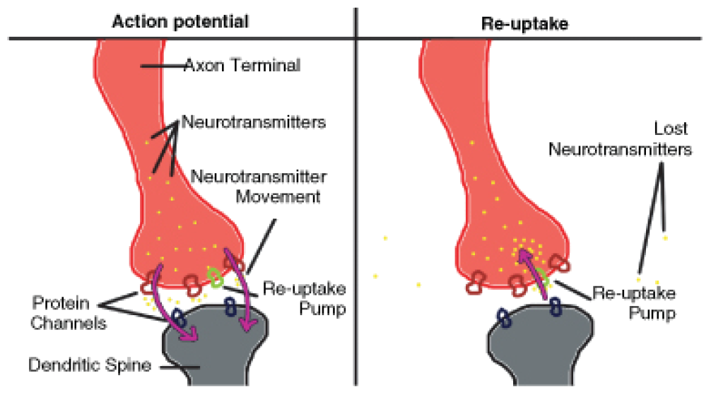
Figure 10: A synapse before and during reuptake. Neurotransmitter transporter proteins (also called reuptake pumps) release the neurotransmitter and also reuptake it from the synaptic cleft. Reuptake is a way of controlling the effect the neurotransmitter has on the post-synaptic cell.
Re-uptake is carried out by transporter proteins which bind to the released transmitter and actively transport it across the plasma membrane into the pre-synaptic neuron. The reuptake of neurotransmitter is the target of some types of medicine. For example, serotonin is a neurotransmitter that is produced by neurons in the brain. Serotonin is believed to into the synaptic cleft, serotonin molecules either attach to the serotonin receptors (called 5-HT receptors) of the post-synaptic neuron, or they attach to receptors on the surface of the presynaptic neuron that produced the serotonin molecules, for reuptake. Reuptake is a form of recycling because the neuron takes back the released neurotransmitter for later use. Medicines called selective serotonin reuptake inhibitors (SSRIs) block the reuptake of the neurotransmitter serotonin. This blocking action increases the amount of serotonin in the synaptic cleft, which prolongs the effect of the serotonin on the postsynaptic neuron. Some scientists hypothesize that decreased levels of serotonin in the brain are linked to clinical depression and other mental illnesses. So SSRI medications such as sertraline and fluoxetine are often prescribed for depression and anxiety disorders.
Another way that a neurotransmitter is removed from a synapse is digestion by an enzyme. At cholinergic synapses (where acetylcholine is the neurotransmitter), the enzyme acetylcholinesterase breaks down the acetylcholine.
Neurotransmitters and Disease
Diseases that affect nerve communication can have serious consequences. A person with Parkinson’s disease has a deficiency of the neurotransmitter dopamine. Progressive death of brain cells that produce dopamine increases this deficit, which causes tremors, and a stiff, unstable posture. L-dopa is a chemical related to dopamine that when given as a medicine, eases some of the symptoms of Parkinson’s disease. The L-dopa acts as a substitute neurotransmitter, but it cannot reverse the disease.
The soil bacterium Clostridium tetani produces a neurotoxin that causes the disease tetanus. The bacteria usually get into the body through an injury caused by an object that is contaminated with C. tetani spores, such as a puncture wound caused by stepping on a nail. The C. tetani neurotoxin blocks the release of the neurotransmitter GABA, which causes skeletal muscles to relax after contraction. When the release of GABA is blocked, the muscle tissue does not relax and remains contracted. Tetanus can be fatal when it affects the muscles used in breathing. Thankfully, tetanus is treatable and can be prevented by vaccination.
Another bacterium called Clostridium botulinum produces a toxin that is occasionally found in preserved foods that have been improperly sterilized. The toxin causes a disease called botulism. Botulin toxin blocks the release of the excitatory neurotransmitter acetylcholine. Blockage of acetylcholine causes the progressive relaxation of muscles because they are unable to contract. Paralysis of the muscles used for breathing can be fatal unless the patient is treated with a respirator.
Synapses and Recent Research
Recent studies have found that electrical synapses are more common in the central nervous system than were previously thought. An electrical synapse is a link between two neighboring neurons that is formed at a narrow gap between the pre- and postsynaptic cells called a gap junction. At gap junctions, cells are about 3.5 nm from each other, a much shorter distance than the 20 to 40 nm distance that separates cells at chemical synapses.

Figure 11: Electrical synapses. Electrical synapses are more common in the nervous system than was once thought. Cell signaling at electrical synapses is much faster than signaling at chemical synapses. The image at the bottom left of the figure shows the location of gap junctions between cells.
Each gap junction has many channels which cross the plasma membranes of both cells, as is shown in Figure 11. Gap junction channels are wide enough to allow ions and even medium sized molecules like signaling molecules to flow from one cell to the next. For example, when positive ions move through the channel into the next cell, the extra positive charges depolarize the postsynaptic cell.
Signaling at electrical synapses is faster than the chemical signaling that occurs across chem chemical messengers, which occurs at chemical synapses. Such fast communication between neurons may indicate that in some parts of the brain large groups of neurons can work as a single unit to process information. Electrical synapses are numerous in the retina and cerebral cortex.
In addition to neurons, glial cells are an important part of the nervous system. The word glia means ”glue” in Greek. Glial cells can be thought of as partners to neurons by aiding in the maintenance of homeostasis, signal transduction, formation of myelin and providing support and nutrition. The importance of neurons as the conductive cells of the nervous system, known as the neuron doctrine, has been questioned by recent research. The role of glial cells in processing neural information has begun to be appreciated more. There are far more glial cells than neurons, it has been estimated that glial cells outnumber neurons by as many as 50:1.
Central Nervous System
The central nervous system (CNS), which includes the brain and the spinal cord, shown in Figure 12, represents the largest part of the nervous system. The brain is the central control of the nervous system. The spinal cord carries nerve impulses from the brain to the body and from the body to the brain. Together with the peripheral nervous system (PNS), which includes all nervous tissue outside of the central nervous system, it controls virtually every activity in the body. The brain is protected by the skull and the spinal cord is protected by the vertebrae.

Figure 12: The components of the central nervous system (CNS).
The Brain
The brain is the most complex organ in the body. The brain contains about 100 billion neurons each of which can be connected to tens of thousands of other neurons within the brain. The brain is the source of what makes us human; the conscious mind. The mind is the set of cognitive processes related to perception, interpretation, imagination, memories, and language. Beyond cognitive functions, the brain regulates processes related to homeostasis such as respiration and heartbeat. An average adult human brain weighs between 1 and 1.5 kg (3 lb). An adult brain uses about 20-25% of the total energy used by the body, while the developing brain of an infant consumes around 60% of total energy used by the body.
The brain can be classified by the processes its different parts control. The cerebrum generally controls conscious functions such as problem-solving and speech, while the midbrain and the brain stem are more involved with unconscious (autonomic) functions such as breathing, heartbeat, and temperature regulation. The cerebellum is involved in coordination and control of body movement.
Cerebrum
The cerebrum is what most people would think of as the ”brain.” The cerebrum lies on top of the brainstem. It is made up of two cerebral hemispheres, which are shown in Figure 13. The two cerebral hemispheres are connected to each other at the corpus callosum, the light-colored X-shaped structure in the center of the image. The corpus callosum is a wide, flat bundle of axons found deep inside the brain. Mammals (including humans), have the largest and most well-developed cerebrum among all species.

Figure 13: A magnetic resonance image (MRI) of the human brain in which the two hemispheres of the cerebrum can be seen.
Each hemisphere of the cerebrum can be divided into four parts, or lobes. These are: the frontal lobe, the parietal lobe, the temporal lobe, and occipital lobe. Researchers have identified a number of functional areas within each lobe, some of which are listed in Table 2. Both hemispheres look identical, but there are functional differences between them. For example, there are differences between the centers of function for spatial awareness between right and left-handed people. Each cerebral hemisphere receives sensory information and controls muscle movements of the opposite side of the body. The right hemisphere controls the left side of the body, and the left hemisphere controls the right side of the body.
Table 2: Functions Controlled by the Cerebral Lobes
| Lobe | Functions |
|---|---|
| Frontal | Speech, intellectual function (reasoning, abstract thought), touch |
| Parietal | Speech, taste, reading |
| Temporal | Hearing, smell |
| Occipital | Vision |
The cerebral cortex is the highly-folded outer layer of the cerebrum that is between 2 mm and 4 mm thick. The lobes that make up the cerebral cortex, shown in Figure 14, are named after the skull bones that cover those areas of the brain. The many folds in the cortex allow for the large surface area of the brain to fit inside the skull. The cerebral cortex controls higher functions, such as consciousness, reasoning, emotions, and language. It also controls sensory functions such as touch, taste, smell, and responses to external stimuli. In the cerebrum, and found below the cerebral cortex, is the white matter. White matter is made up of myelinated axons that act as “cables” that link up certain parts of the right and left hemispheres.

Figure 14: The lobes of the cerebral cortex-frontal, temporal, occipital, and parietal. The cerebellum (purple) and brain stem (gray) are not part of the hindbrain. In vertebrates, a gross division into three major parts is used.
Diencephalon
The diencephalon is the region of the brain that includes structures such as the thalamus, the hypothalamus, and a portion of the pituitary gland. The thalamus is believed to “translate” sensory signals for the cerebral cortex. The thalamus also plays an important role in regulating states of sleep and wakefulness. The hypothalamus gland controls certain metabolic processes and other autonomic activities such as body temperature, hunger, thirst, and circadian cycles. The hypothalamus also makes and releases neurotransmitters that control the action of the pituitary gland. The thalamus, hypothalamus, and hippocampus together are considered part of a set of structures called the limbic system. The limbic system is considered the “emotional center” of the brain.
Brain Stem
Sometimes called the “lower brain,” the brain stem is the lower part of the brain that is joined to the spinal cord. There are three parts to the brainstem: the midbrain, the pons, and the medulla oblongata, shown in Figure 15. The midbrain is more involved with unconscious, autonomic functions. The midbrain deals with several types of sensory information including sound and sight. It also “translates” sensory information to be sent to the forebrain. The brainstem also helps coordinate large body movements such as walking and running. The pons relays messages to different parts of the brain (the cerebrum and cerebellum), and helps regulate breathing. Some researchers propose that it has a role in dreaming. The medulla oblongata, also called the medulla, shares some of the function of the pons. It controls several homeostatic functions that you are usually unaware of, such as breathing, heart and blood vessel activity, swallowing, and digestion.

Figure 15: The locations of the brainstem and cerebellum. The brainstem is in the center of this image. It is made up of the pons, medulla oblongata, and the midbrain. The cerebellum is the red structure to the right of the brainstem.
One of the brain stem’s most important roles is that of an “information highway.” That is, all of the information coming from the body to the brain (sensory) and the information from the cerebrum to the body (motor) go through the brain stem. Sensory pathways for such things as pain, temperature, touch, and pressure sensation go upward to the cerebrum, and motor pathways for movement and other body processes go downward to the spinal cord. Most of the axons in the motor pathway cross from one side of the CNS to the other as they pass through the medulla oblongata. As a result, the right side of the brain controls much of the movement in the left side of the body, and the left side of the brain controls much of the movement in the right side of the body.
Cerebellum
The cerebellum is found just below the occipital lobe of the cerebrum. It plays an important role in coordination and the control of body movements. Many nerve pathways link the cerebellum with motor neurons, which are neurons that send information to the muscles causing them to move, and a group of nerves that provides information on the position of the body in space. The cerebellum processes information from both these pathways, and uses the feedback on body position to fine-tune body movements. Hand-eye coordination is an example of such a function. If the cerebellum is damaged, there will not be paralysis, but the fine movement of the body (such as hand-eye coordination), balance, posture, and the ability to learn new motor skills will be negatively affected. The cerebellum is the purple structure in Figure 14. A section of the cerebellum is shown in Figure 15.
Spinal Cord
The spinal cord is a thin, tubular bundle of nervous tissue that extends from the medulla oblongata and continues to the lower back, where it ends in a group of fibrous extensions. It is protected by the spinal vertebrae. The main function of the spinal cord is as an information superhighway that links the sensory messages from the body to the brain. The outer cortex of the cord contains white matter (myelinated sensory and motor neurons). The central region, the grey matter, is made up of unmyelinated neurons. A cross section of the spinal cord is shown in Figure 16.

Figure 16: A cross section of the spinal cord. The central butterfly-shaped area is the gray matter and the area that surrounds it is the outer cortex (made up of white matter). Instructions go to the body’s muscles and other areas through the motor neurons that leave the spinal cord in the spinal nerves. Sensory information from the body enters the spinal cord through sensory neurons.
Peripheral Nervous System
The peripheral nervous system (PNS) consists of the nervous tissue that lies outside the central nervous system, shown in Figure 17. The nervous tissue of the peripheral nervous system serves the limbs and organs. The central nervous system interacts with the peripheral nervous system through twelve pairs of cranial nerves that connect the brain to areas of the head and neck and 31 pairs of spinal nerves that connect the spinal cord (and CNS) to the rest of the body, such as the internal organs, arms, and legs. A nerve is an enclosed, cable-like bundle of axons. Unlike the central nervous system, the peripheral nervous system is not protected by bone, making it more vulnerable to toxins and injuries.

Figure 17: The peripheral nervous system (PNS). The peripheral nervous system extends from the CNS and reaches out to all parts of the body, from the cranial nerves found in the head to the plantar nerves in the tips of the toes.
Spinal nerves originate from the spinal cord. They control functions of the rest of the body. Each spinal nerve has a dorsal root and a ventral root, which are shown in Figure 18. The dorsal root is the “nerve highway” that carries sensory information from sensory receptors in the body to the CNS. The ventral root contains axons of motor neurons which carry information away from the CNS to the muscles and glands of the body.

Figure 18: A cross section of the spinal cord. The central butterfly-shaped area (1, 2, 3) is the gray matter, the outer cortex is the white matter. Instructions going to the body’s muscles and other areas go through the motor neurons that leave the spinal cord in the ventral roots (11). Sensory information from the body enters the spinal cord through sensory neurons in the dorsal roots (12). Dorsal and ventral roots occur on both sides of the spinal cord, only one side is shown in this diagram.
These two nerve “highways” are actually parts of two subdivisions of the PNS. The sensory division, also known as the afferent division, carries sensory information from sensory receptors in the body to the CNS. The sensory division keeps the CNS constantly updated on events happening inside and outside the body. The motor division, or efferent division, carries nerve impulses from the CNS to the muscles, glands and organs of the body. The nerve impulses of the motor division cause muscles to contract and cause glands to secrete chemical signals.
Somatic and Autonomic Nervous Systems
The motor division of the peripheral nervous system is divided into the somatic nervous system and the autonomic nervous system:
The somatic nervous system is the part of the PNS that is associated with the conscious (voluntary) control of the body through the movement of skeletal muscles and the perception of external stimuli through senses such as touch, hearing, and sight. The system includes all the neurons connected with muscles, skin and sense organs. The somatic nervous system is made up of sensory nerves that receive sensory information from the external environment, and motor nerves responsible for muscle contraction.
Together with interneurons, the sensory and motor neurons are found in a reflex arc. A reflex is an automatic (involuntary) action caused by a defined stimulus and carried out through a reflex arc. For example, a person stepping on a sharp object would start the reflex action through the creation of a stimulus, (pain) within specialized pain receptors located in the skin tissue of the foot. The resulting stimulus would be passed along sensory neurons to the spinal cord. This stimulus is usually processed by an interneuron to create an immediate response to pain by initiating a motor response in the muscles of the leg which pull the foot away from the object. This reflexive action would occur as the pain sensation is arriving in the brain. A reflex arc is shown in Figure 19.

Figure 19: The components of a reflex. A sensory receptor that detects a stimulus and sends nerve signals to the spinal cord. These signals activate motor neurons that lead back to the effector (muscle).
The autonomic nervous system (ANS) is the part of the peripheral nervous system that maintains homeostasis in the body. Your body carries out most of these maintenance activities without your conscious control, which is why the autonomic nervous system is also called the involuntary nervous system. The ANS has far reaching effects, such as the control of heart rate, digestion, respiration rate, salivation, and perspiration. Some autonomic nervous system functions work in line with the conscious mind, such as breathing.
The ANS is also made up of the sensory and motor neurons that send messages to and from the internal organs. These neurons form reflex arcs that pass through the medulla oblongata. This explains why even a person’s cerebrum may experience trauma, yet their cardiovascular, digestive and respiratory functions will continue even if higher level functions such as awareness and consciousness, are lost. Such a low level of brain functioning is referred to as a vegetative state.

Figure 20: Watch out! A situation in which your sympathetic nervous system (and hopefully your somatic nervous system), would be firing at full speed.
The ANS has two subdivisions: the sympathetic division and parasympathetic division. The sympathetic division generally stimulates body systems during emergency situations. It gets the body ready for ”fight or flight,” which would probably be required by the situation shown in Figure 20, while the parasympathetic division controls non-emergency functions such as digestion. The relationship between the divisions of the nervous system is illustrated in Figure 21.
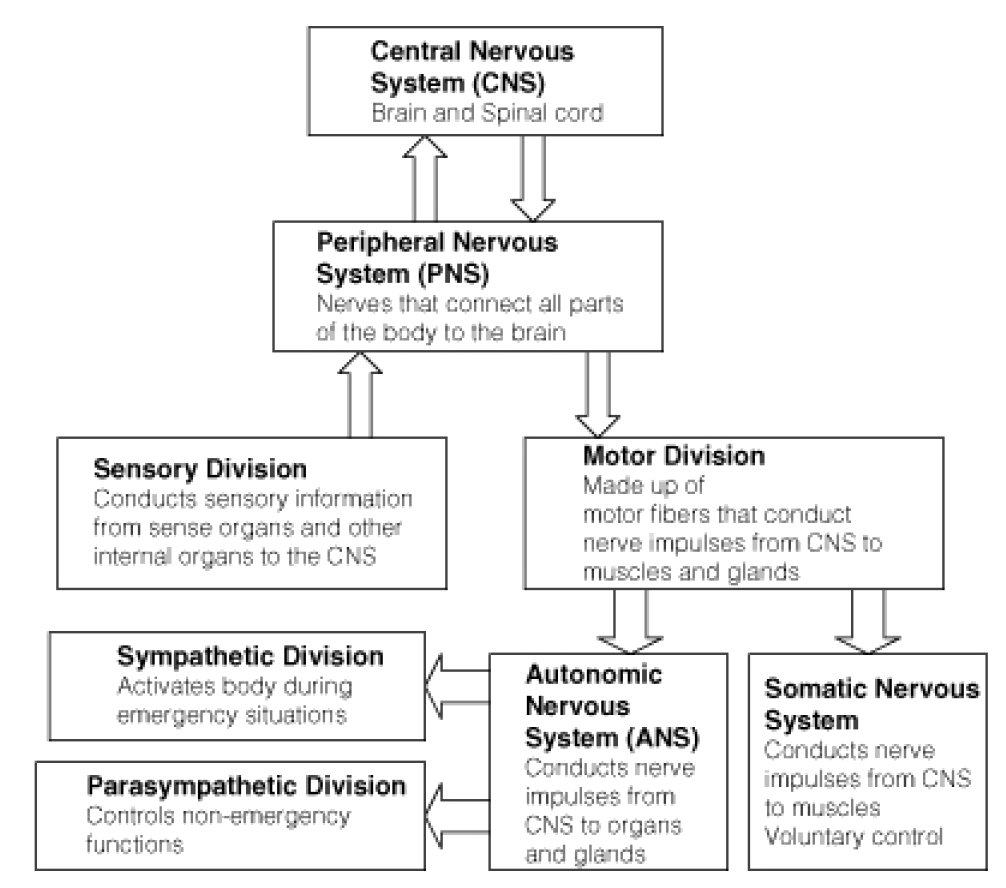
Figure 21: Levels of Organization of the Nervous System.
Sense Organs and Sensory Perception
Your senses are your body’s means of making sense of the information your nervous system receives from inside your body and from the outside world. Your senses enable you to adapt to change in your environment and survive. The sensory division of the peripheral nervous system is organized into highly developed sense organs, which are groups of tissues that work together in responding to a specific kind of physical stimulus, such as the stimulus in Figure 22. The sense organs correspond to a defined region (or group of regions) within the brain where the nerve signals are received and interpreted. Your sense organs include your eyes, ears, nose, mouth, and skin. They all have sensory receptors that are specific for certain stimuli. For example, the nose has sensory receptors for odors (smells). Sensory neurons send nerve impulses from sensory receptors to the central nervous system. The brain then interprets the nerve impulses to form a response.

Figure 22: Can you smell these fresh, juicy oranges and kumquats? Your senses of smell, taste and sight are also important in developing an appetite. Just think of how appetizing these fruits would be if they were blue, crunchy, and smelled like burned toast.
A sensory receptor is a cell, or a group of cells that detect stimuli. Sensory receptors can be classified based on the type of stimuli to which they respond.
• Chemoreceptors respond to chemical stimuli.
• Mechanoreceptors respond to mechanical stress or strain (movement).
• Thermoreceptors respond to temperature changes.
• Photoreceptors respond to variations in light.
• Baroreceptors respond to pressure.
Specific areas of the brain interpret information from each sense organ. For example, regions of the occipital lobe interpret nerve impulses that come from the sensory receptors of the eyes, and regions of the temporal lobe interpret sensory information from the ears through the nerves that enter the brain in these areas, as shown in Figure 23. It is generally agreed that there are at least seven different senses in humans. These are sight, sound, taste, smell, touch, balance, and body awareness (the sense of knowing where the regions of your body are located at any one time). At least two other senses that humans do not have are observed in other organisms. Examples include electroreception, the ability to detect electric fields, and magnetoreception, the ability to detect magnetic fields.

Figure 23: The entry of sensory nerves into the brain. Among other nerves, the sensory nerves for smell, sight, hearing, and taste (yellow structures) can be seen entering the skull. You can also see how the cerebrum, thalamus, cerebellum, and brain stem are nested within the skull.
Sight
Sight or vision describes the ability of the brain and eye to detect certain wavelengths of electromagnetic radiation (light), and interpret the image as ”sight.” Different receptors are responsible for the perception of color (the frequency of photons of light) and perception of brightness (number of photons of light). Photoreceptors are found in the retina, shown in Figure 24.

Figure 24: The structure of the eye. The macula is a spot near the center of the retina that has a diameter of about 1.5 mm. Near its center is the fovea, a small pit that contains the largest concentration of cone cells in the eye and is responsible for central vision. The macula is the point of sharpest vision.
The structure of the eye owes itself completely to the task of focusing light onto the retina, the light-sensitive inner layer of the eye. First, light passes through a clear protective layer called the cornea, shown in Figure 24. Light then passes through the pupil, which is the opening in the iris, and into the interior of the eye. After passing through the pupil, the light then travels through the lens, a transparent, biconvex structure that, along with the cornea, helps to focus light on the retina. Muscles attached to the lens change the shape of the lens to bend the light rays so that they focus on the retina, as shown in Figure 25. Light hitting the retina causes chemical changes in the photosensitive cells of the retina, the products of which trigger nerve impulses which travel to the brain along the optic nerve.

Figure 25: Focusing light in the retina. This diagram shows how light from a distant source is bent by the stretched lens to strike the retina, and how light from a closer source is bent even more sharply by the relaxed lens to strike the retina.
The retina has two forms of photosensitive cells important to vision—rods and cones. Rod cells are highly sensitive to light which allows them to respond in dim light and dark conditions, but, they cannot detect color. These are the cells which allow humans and other animals to see by moonlight, or in a dimly-lit room. This is why the darker conditions become, the less color objects seem to have. Cone cells respond to different wavelengths of bright light to initiate a nerve impulse. They are also responsible for the sharpness of images. Cones do not respond well in poor light conditions, which is the reason why you see things in dim light as fuzzy shades of gray.
Humans have three different types of cone cells that respond to different wavelengths of light. These cone cells contain a pigment that absorbs the energy from different wavelengths of light to initiate a nerve impulse. Activation of the visual pigments by certain wavelengths of light opens ion channels on the membrane of the cone or rod cell. This leads to an action potential that is carried by the millions of neuron axons that make up the optic nerve to the visual centers of the brain. The brain integrates the nerve impulses from the cone cells and perceives the world in all the colors of the visual spectrum. A person who is colorblind has damaged or missing cones, and is unable to perceive certain colors.
Hearing
Hearing is the sense of sound perception that results from the movement of tiny hair fibers in the inner ear. These hairs detect the motion of a membrane which vibrates in response to changes in air pressure. Sound can also be detected as vibrations that are conducted through the body. Sound wave frequencies that are too low or too high to be heard by the ear can be detected this way. Audible sound is sensed by the ear.
The folds of cartilage surrounding the outer ear canal are called the pinna. Sound waves are gathered by the pinna, and channeled down the auditory canal, a tube-shaped opening of the ear which ends at the tympanic membrane, or eardrum.
Sound waves traveling through the ear canal hit the eardrum and cause it to vibrate. This wave information travels across the air-filled middle ear cavity through a group of three tiny, delicate bones: the hammer, the anvil, and the stirrup, shown in Figure 26. This group of bones transfers the eardrum vibrations to another membrane called the oval window. The oval window separates the middle ear from the inner ear. The inner ear contains the cochlea.

Figure 26: The detection of sound by your ear.
The cochlea is a coiled tube that is filled with a watery liquid, which moves in response to the vibrations coming from the middle ear through the oval window. As the fluid moves, thousands of mechanoreceptors called hair cells bend, releasing a neurotransmitter. The neurotransmitter causes an action potential in the neurons of the auditory nerve. The action potential travels along the auditory nerve to structures in the brainstem, then to the thalamus, and then to the auditory centers of the brain in the temporal lobe of the cerebral cortex.
A very strong movement of the fluid within the cochlea, caused by very loud noise, can kill hair cells. This is a common cause of partial hearing loss and is the reason why users of firearms or heavy machinery should wear earmuffs or earplugs. Destruction of the hair cells usually leads to permanent hearing loss because once destroyed, the hairs do not generally grow back.
Balance and the Ears
It might be hard to believe, but your ears are also in charge of your sense of balance! The semicircular canals are three fluid-filled interconnected tubes found inside each ear. They can be seen in Figure 26, directly above the cochlea. The canals are positioned at angles between 95 to 115 degrees relative to one another. The angles between the canals are not perpendicular, so movements of the head cause movement of fluid in two canals at the same time.
Each canal is filled with fluid called endolymph and motion sensors with little hairs, called cilia, line each canal. Movement of the head and body cause the endolymph in the canals to move about. The hair cells sense the strength and direction of the fluid’s movement and send electrical signals to the cerebellum which interprets the information and responds to help keep the body’s sense of balance. The interaction of the semicircular canals and the cerebellum allow the performer in Figure 27 to do his act.

Figure 27: Good balance required! This performer’s sense of balance is dependent on communication between his semicircular canals and his cerebellum.
When the sense of balance is interrupted it causes dizziness and nausea. Balance can be upset by an inner ear infection, a bad head cold or a sinus infection, or a number of other medical conditions. It can also be temporarily disturbed by rapid and repetitive movement, for example riding on a merry-go-round or spinning around in a circle.
Taste and Smell
Taste is one of the two main chemical senses, the other being smell. There are at least four types of taste receptors on the tongue. Taste stimuli from each receptor type send information to a different region of the brain. The four well-known receptors detect sweet, salt, sour, and bitter. The existence of a fifth receptor, for a sensation called umami, was confirmed in 2000. The umami receptor detects the amino acid glutamate, which causes a savory, “meaty” flavor in foods.
The chemoreceptors of the mouth are the taste cells that are found in bundles called taste buds. Most of the taste buds are embedded within the tiny papillae or “bumps” that cover the tongue, shown in Figure 28. Each receptor has a different way of detecting certain compounds and starting an action potential which alerts the brain. The compounds bind to receptors in the taste cells and stimulate neurons in the taste buds. The action potential moves along the facial nerves to the thalamus and then to the taste center of cerebral cortex for interpretation by the brain. The tongue can also feel sensations that are not generally called tastes. These include: temperature (hot or cold), coolness (as in “minty” or “fresh”), spiciness or hotness (peppery), and fattiness (greasy).

Figure 28: The location of taste buds. Most of the taste buds in the mouth are embedded in the papillae, the little bumps that cover the tongue. The deep groove (fissure) that runs down the center of the tongue in this photo is a common and perfectly normal condition.
Smell is the other ”chemical” sense. The chemoreceptors of smell are called olfactory receptors. About 40 million olfactory receptor neurons line the nasal passages. Different odor molecules bind to and excite specific olfactory receptors. The combination of excitatory signals from different receptors makes up what we identify as “smell.” Signals from the olfactory receptors travel along nerves to the olfactory bulb in the brain where they then move to the smell center in the frontal lobe of the cerebral cortex. Olfactory receptor neurons in the nose differ from most other neurons in that they die and regenerate on a regular basis. A dog’s keen sense of smell is due to the large area of its nasal passages that are covered by olfactory receptors, and the large number of nerves that bring nerve impulses from the receptors to its brain. For example, the area in which olfactory receptors are located inside the human nose (called the olfactory epithelium), which is shown in Figure 29, measures about 12 \(cm^2\). The olfactory epithelium of some dogs’ noses can measure about 150 \(cm^2\)!

Figure 29: The location of olfactory nerves. Olfactory receptors and their associated nerves (yellow) line the top of the nasal passages. Nerve messages from the receptors are sent to the brain to be interpreted as certain smells.
Have you ever noticed that you cannot taste anything when your nose is stuffed up? That is because your senses of smell and taste are closely linked. This is due to the fact that your nasal cavity, located behind the nostrils, connects to your mouth at the back of your throat, as shown in Figure 29. Your olfactory receptors and taste receptors both contribute to the flavor of food. Your tongue can only tell among a few different types of taste, while your nose can distinguish among hundreds of smells, even if only in tiny amounts.
Touch, Pressure, and Pain
Touch is the sense of pressure perception, which is generally felt in the skin. There are a variety of pressure receptors that respond to variations in pressure and tension. Mechanoreceptors are most numerous on the tongue, lips, face, palms (including fingertips), and soles of the feet.
There are several types of pain receptors, called nociceptors, which respond to potentially damaging stimuli. They are mostly found in the external parts of the body such as the skin, cornea, and mucous membranes, but are also found in muscles, joints, and some internal organs. Nociceptors are classified according to the stimuli to which they respond: thermal, mechanical or chemical. But some receptors respond to many different damaging stimuli of a chemical, thermal, or mechanical nature. Thermal receptors are activated by potentially harmful heat or cold, temperatures above 45°C and below 5°C. Mechanical receptors respond to excess pressure, squeezing, or bending, the type of painful stimuli that a cactus such as the one in Figure 30 would cause. Together these nociceptors allow the organism to feel pain in response to damaging pressure, excessive heat, excessive cold and a range of chemicals, the majority of which are damaging to the tissue surrounding the nociceptor.

Figure 30: Mechanical pain receptors in your skin would warn you if you got too close to this prickly cactus.
Drugs and the Nervous System
A drug is any chemical or biological substance that affects the body’s structure or functions. Drugs in the form of medicines are used to treat many illnesses and disorders. A medicine (or medication), is a drug that is taken to cure or reduce the symptoms of an illness. However, drugs, whether they are medicines, legal or illegal drugs, can be abused for the effects they have on the central nervous system (CNS). In fact many medical uses of drugs depend on the powerful effect they have on brain function. For example, anti-depression medicines are used to treat depression and anxiety disorders, and antipsychotic medicines are used to treat schizophrenia and bipolar disorder.
A psychoactive drug is a substance that affects the central nervous system by altering cognitive function. Change in cognitive function results in changes in how a person feels, thinks, perceives, and acts. Almost everyone has used a psychoactive drug at some time in their life, and many people take such drugs daily. For example, the coffee or tea that you may have drank to waken yourself up this morning, or the cola, energy drink, or chocolate that you had as a snack contain the psychoactive drug caffeine. Caffeine is a CNS stimulant that makes you feel less drowsy and more alert. Coffee beans, the most common source of caffeine, are shown in Figure 31.

Figure 31: Roasted coffee beans. Coffee beans are a common source of the stimulant caffeine. Other plant sources include the leaves of tea, cocoa, yerba mate, and guarana plants. These plants use caffeine as a means of protection against being eaten. The caffeine in the leaves of these plants can paralyze and kill the insects that feed upon them.
Drugs and the Brain: How Psychoactive Drugs Work
How we perceive stimuli, feel, think, and do is a result of neurons sending action potentials and neurotransmitters to each other and to other cells in the body. Psychoactive drugs affect how neurons communicate with each other. These drug molecules can alter neurotransmission, by blocking receptor proteins, mimicking neurotransmitters, or changing the amount of neurotransmitter in the synapse, shown in Figure 32, by blocking reuptake. In this way a psychoactive drug can change how we feel, think, and interact with the world. Sometimes such effects are beneficial, such as taking a prescribed painkiller (hydrocodone, for example), to ease the pain of a broken bone. Sometimes the effects are harmful, which could happen if the person continued to take the powerful painkiller long after their broken bone had healed. Some examples of psychoactive medicines are listed in Table 3.

Figure 32: The release of neurotransmitter into the synaptic cleft. Depending on its method of action, a psychoactive substance may block the receptors on the post-synaptic neuron, or block reuptake or affect neurotransmitter synthesis in the pre-synaptic neuron.
Table 3: Some Psychoactive Medicines and Their Uses
| Type | Uses | Example | Action |
|---|---|---|---|
| Anesthetics | Block pain and other sensations. Often induce unconsciousness, which allows patients to undergo medical procedures. | Lidocaine, nitrous oxide | Mimic the inhibitory neurotransmitter GABA, or increase the amount of GABA in the synapse which prevents an action potential. |
| Painkillers (analgesics) | Reduce the sensation of pain. Includes narcotics and non-steroidal anti-inflammatory drugs (NSAIDS) | Narcotics: morphine and codeine NSAIDS: aspirin and acetaminophen (paracetamol). | Drug molecules mimic endogenous opioids “natural painkillers,” such as endorphins, by binding to opioid receptors. |
| Antidepressants | Antidepressants are used to treat disorders such as clinical depression, anxiety, and eating disorders | Selective Serotonin Reuptake Inhibitors (SSRIs); Monoamine oxidase inhibitors (MAOIs) | SSRIs: Block the uptake of the neurotransmitter serotonin by presynaptic neuron. MAOIs: Prevent an enzyme from breaking down serotonin in the synapse. Both actions result in an increase of serotonin in the synapse. |
| Stimulants | Used to treat disorders such as attention deficit disorder and to suppress the appetite | Amphetamine salts | Increases extracellular levels of dopamine, norepinephrine and serotonin by various means |
| Antipsychotics | Used to treat psychoses such as schizophrenia and mania. | Chlorpromazine | Blocks dopamine receptors in post synaptic neurons |
| Cough medicines (antitussives) | Used to treat persistent coughing. | Dextromethorphan (DXM) and codeine | Inhibit the action of, the NMDA receptor in the post synaptic cell. Reduces action potential, similar in action to anesthetics |
Drug Abuse
Psychoactive drugs bring about changes in mood and feelings that a user may find desirable, therefore many psychoactive substances are abused. Drug abuse is the repeated use of a drug without advice or guidance of a medical professional, and use for reasons other than for what the drug was originally intended. With continued use of a drug, a person might find that they cannot function normally without the drug, a state called physical dependence. However, note that physical dependence is not in itself bad, for example, a person who has diabetes is physically dependent on insulin injections. Their body cannot work properly without it. Emotionally or mentally needing a drug to be able to function normally is called psychological dependence. When a person continues to take a psychoactive drug, they eventually need to take larger doses of the drug to get the desired effect; this process is known as building a tolerance to the drug. Drug tolerance can involve both psychological and physical factors.
A person who is abusing a drug may eventually lose control of their drug-taking behavior, partly due to the changes the drug has caused in their brain, and partly due to learned drug-abuse behaviors (such as stealing and lying to get money or drugs). In the state of addiction, a drug addict’s life and activities revolve around getting more of the drug to feed their habit, even if it leads to severe consequences such as getting arrested, dropping out of school, or isolation from friends and family. In a person who is addicted to a drug, the pattern of increasing dose due to tolerance can lead to a drug overdose, also known as an OD. A drug overdose is generally considered harmful and may lead to death. Drug dependence and addiction are caused by changes in the way neurons in the CNS send and receive neurotransmitters. It is for this reason dependency and addiction are treated as brain disorders by medical professionals.
Several classes of psychoactive drugs are commonly abused. Stimulants such as cocaine, nicotine, and amphetamine increase the activity of the sympathetic nervous system, the central nervous system, or both. Stimulants generally increase heart rate, blood pressure, and increase the sense of alertness. Some stimulants, such as caffeine, are used medicinally to increase or maintain alertness, and to counteract fatigue. High doses of stimulants can be fatal. A common source of nicotine is cigarette tobacco, shown in Figure 33.

Figure 33: Cigarettes are a common source of nicotine. Nicotine is a compound that is found in the leaves of the tobacco plant. It is a potent neurotoxin for insects, and was once used as an insecticide. In addition to the addictive nature of nicotine, long-term tobacco use carries significant risks of developing various cancers as well as strokes and severe cardiovascular and respiratory diseases.
Hypnotics, also known as depressants, such as alcohol, codeine, barbiturates, and benzodiazepines generally decrease the activity of the central nervous system. Depressants slow down brain function and give a drowsy or calm feeling. However, taking too much of a depressant drug can cause dangerously slow breathing and heart rates, and may result in death. Many depressants acting on the CNS do so by increasing the activity of the inhibitory neurotransmitter gamma-aminobutyric acid (GABA), although there are many receptors that are affected by different depressants. GABA calms the activity of the CNS and promotes sleep. Drugs that stimulate the activity of this amino acid slow down brain function and cause a drowsy or calm feeling, so depressants are generally prescribed to relieve symptoms of anxiety or insomnia.
Hallucinogens, also known as psychedelic drugs, such as lysergic acid diethylamide (LSD), phencyclidine (PCP), and ketamine, are psychoactive drugs that do not increase or decrease a certain feeling or emotion, but rather they induce experiences, such as sensory distortions and “out-of-body experiences,” that are very different from those of ordinary consciousness. These experiences are often called trance-like states. The use of psychedelic drugs has been linked to a potential for brain damage.
There are many ways in which psychoactive drugs can affect the CNS. Each drug has a specific action on one or more neurotransmitters or receptors. Drugs that increase activity in particular neurotransmitter systems are called agonists. They act by increasing the synthesis of one or more neurotransmitters or reducing its reuptake from the synapses. Drugs that reduce neurotransmitter activity are called antagonists, and work by interfering with synthesis or blocking postsynaptic receptors so that neurotransmitters cannot bind to them. The drug ketamine, which is used as an anesthetic and a painkiller, blocks the action of the neurotransmitter glutamate. Diacetylmorphine (heroin) enhances the action of endorphins in the brain. Different drugs also affect different parts of the brain. For example, drugs that affect breathing, such as cough suppressants, affect the brainstem to stop the coughing reflex. Painkillers (analgesics) block pain messages coming through the spinal cord from the body. In Figure 34 the brain-stem region is blue, and the spinal cord is yellow.

Figure 34: The limbic system (in red) includes structures in the human brain that have been linked to emotion, motivation, and emotional association with memory. The action of neurotransmitters in the limbic system is altered by addictive drugs.
How Addiction Happens
The neurobiological theory of addiction proposes that certain chemical pathways are greatly changed in the brain of an addicted person. Almost all drugs that are abused affect a certain set of brain structures in the limbic system called the ”brain reward system,” shown in Figure 27. The neurotransmitter dopamine is commonly associated with the brain reward system. The system providing feelings of pleasure (the “reward”), that motivates a person to perform certain activities over and over again. Dopamine is released at synapses by neurons when a person has a pleasurable experience such as eating a favorite food, or eating when very hungry. Such mechanisms have evolved to ensure the survival of organisms.
Some drugs, such as cocaine, nicotine, amphetamines, and alcohol directly or indirectly increase the amount of dopamine in the limbic structures. The pleasurable feelings that these drugs produce trick the body into thinking that the drug is good,important for survival, and needs to be taken repeatedly. Drugs that directly affect the brain reward system are highly addictive. The stimulant nicotine, which is found in tobacco, is highly addictive.
Cocaine is an example of a psychoactive drug that is both used as a medicine, and abused as a drug. Cocaine is highly addictive. It is a dopamine transporter blocker—it blocks the reuptake of dopamine by the presynaptic neuron. This action increases the amount of dopamine left in the synaptic cleft, so dopamine has a stronger effect on the postsynaptic neuron. Continued cocaine use causes a reduction in the number of dopamine receptors on the postsynaptic neuron. Eventually, the post synaptic neuron becomes understimulated because there are fewer dopamine receptors on it to respond to dopamine. At this point, more cocaine must be taken to stimulate the postsynaptic neuron into an action potential. If a person becomes dependent on the drug, they need cocaine for their body to act normally.
If a person were to stop taking the drug at this point, their body would not be able to act normally, and they would experience a range of uncomfortable and painful symptoms called withdrawal. Symptoms of withdrawal include vomiting, diarrhea, and depression.
Many psychoactive substances are used or abused for their mood and perception altering effects, including those with accepted uses in medicine and psychiatry. Classes of drugs that are frequently abused include some of the drugs listed in Table 4. Drugs that are deemed by to have no medical uses and a high potential for abuse are usually illegal.
Not all drugs are physically addictive, but any activity that stimulates the brain reward system can lead to psychological addiction. Drugs that are most likely to cause addiction are drugs that directly stimulate the dopaminergic system, like cocaine, nicotine, and amphetamines. Drugs that only indirectly stimulate the dopaminergic system, such as psychedelics, are not as likely to be addictive.
Table 4: Some Common Drugs of Abuse
| Psychoactive Drugs | Effects | Example | Some Common Forms or Names |
|---|---|---|---|
| Stimulants | Elevate the central nervous system and raise level of alertness and wakefulness | Caffeine, cocaine, amphetamine, methamphetamine | Coffee, coke, meth, ecstasy (X) |
| Hallucinogens | Induce perceptual and cognitive distortions | LSD, psilocybin, mescaline, PCP | Acid, magic mushrooms, peyote, angel dust |
| Hypnotics | Depress the CNS, and induce sleep | Barbiturates, opioids (e.g. codeine, morphine, oxycodone), benzodiazepines, ethanol | Diazapam, alcohol |
| Analgesics | Induce euphoria, reduce sensation of pain | Codeine, morphine, ketamine, heroin, phencyclidine (PCP), tetrahydrocannabinol (THC) | Horse, angel dust, cannabis, marijuana |
Images courtesy of:
http://www.flickr.com/photos/bike/248030261/. CC-BY-SA 2.0.
http://commons.wikimedia.org/wiki/Image:Neuron.svg. GNU-FDL 1.2.
http://commons.wikimedia.org/wiki/File: Complete_neuron_cell_diagram_en.svg. CC-BY-SA.
http://commons.wikimedia.org/wiki/File: Complete_neuron_cell_diagram_en.svg. Public Domain.
http://commons.wikimedia.org/wiki/Image:IonChannels.jpg. Public Domain.
http://en.wikipedia.org/wiki/Image:Action_potential_vert.png. GNU-FDL.
http://commons.wikimedia.org/wiki/Image:Synapse_diag3.png. CC-BY-SA.
http://commons.wikimedia.org/wiki/Image:Synapse_diag4.png. GNU-FDL.
http://en.wikipedia.org/wiki/Image:Reuptake_both.png. Public Domain.
LadyofHats. http://en.wikipedia.org/wiki/File:Gap_cell_junction_en.svg. Public Domain.
http://en.wikipedia.org/wiki/Image:Central_nervous_system.svg#en. Public Domain.
http://en.wikipedia.org/wiki/Image:User-FastFission-brain-frame44.png. GNU-FDL.
http://en.wikipedia.org/wiki/Image:Brain_lateral_view.svg. CC-BY-SA-2.5.
Gray’s Anatomy. http://en.wikipedia.org/wiki/Image:Gray720.png. Public Domain.
http://commons.wikimedia.org/wiki/Image:Gray770.png. Public Domain.
http://commons.wikimedia.org/wiki/Image:Nervous_system_diagram.png. Public Domain.
http://en.wikipedia.org/wiki/Image:Medulla_spinalis_-Section-_English.svg. CC-BY-SA.
http://www.flickr.com/photos/pete/13059185/. CC-BY-SA-2.0.
http://www.flickr.com/photos/laurelfan/765440395/. CC-BY-SA.
http://commons.wikimedia.org/wiki/Image:Skull_and_brainstem_inner_ear.svg. CC-BY 2.5.
http://en.wikipedia.org/wiki/Image:Focus_in_an_eye.svg. CC-BY-SA 2.5.
http://www.flickr.com/photos/hourann/454064419/. CC-BY-SA.
http://en.wikipedia.org/wiki/Image:Tongue.agr.jpg. GNU-FDL.
http://commons.wikimedia.org/wiki/Image:Head_olfactory_nerve.jpg. CC-BY 2.5.
http://en.wikipedia.org/wiki/Image:Human_eye_cross-sectional_view_grayscale.png. Public Domain.
http://www.flickr.com/photos/valeriebb/295872714/. CC-BY 2.0.
http://www.flickr.com/photos/abdelazer/528578931/. CC-BY-SA 2.0.
http://en.wikipedia.org/wiki/Image:SynapseIllustration2.png. CC-BY-SA.
http://www.flickr.com/photos/crucifixioncruise/1195949382/. CC-SA-BY.
http://en.wikipedia.org/wiki/Image:Brain_limbicsystem.jpg. Public Domain.