Introduction to Cells
Article objectives
Knowing the make up of cells and how cells work is necessary to all of the biological sciences. Learning about the similarities and differences between cell types is particularly important to the fields of cell biology and molecular biology. The importance of the similarities and differences between cell types is a unifying theme in biology. They allow the principles learned from studying one cell type to be applied when learning about other cell types. For example, learning about how single-celled animals or bacteria work can help us understand more about how human cells work. Research in cell biology is closely linked to genetics, biochemistry, molecular biology, and developmental biology.
Discovery of Cells
A cell is the smallest unit that can carry out the processes of life. It is the basic unit of all living things, and all organisms are made up of one or more cells. In addition to having the same basic structure, all cells carry out similar life processes. These include transport of materials, obtaining and using energy, waste disposal, replication, and responding to their environment.
If you look at living organisms under a microscope you will see they are made up of cells. The word cell was first used by Robert Hooke, a British biologist and early microscopist. Hooke looked at thin slices of cork under a microscope. The structure he saw looked like a honeycomb as it was made up of many tiny units. Hooke’s drawing is shown in Figure 1. In 1665 Hooke published his book Micrographia, in which he wrote:
... I could exceedingly plainly perceive it to be all perforated and porous, much like a Honey-comb, but that the pores of it were not regular.... these pores, or cells, ... were indeed the first microscopical pores I ever saw, and perhaps, that were ever seen, for I had not met with any Writer or Person, that had made any mention of them before this...

Figure 1: Drawing of the structure of cork from Micrographia as it appeared under the microscope to Robert Hooke. The first scientific use of the word cell appears in this book.
During the 1670s, the Dutch tradesman Antony van Leeuwenhoek, shown in Figure 2, used microscopes to observe many microbes and body cells. Leeuwenhoek developed an interest in microscopy and ground his own lenses to make simple microscopes. Compound microscopes, which are microscopes that use more than one lens, had been invented around 1595. Several people, including Robert Hooke, had built compound microscopes and were making important discoveries with them during Leeuwenhoek’s time. These compound microscopes were very similar to the microscopes in use today. However, Leeuwenhoek was so good at making lenses that his simple microscopes were able to magnify much more clearly than the compound microscopes of his day. His microscope’s increased ability to magnify over 200 times is comparable to a modern compound light microscope.

Figure 2: Antony van Leeuwenhoek (1632-1723). His carefully crafted microscopes and insightful observations of microbes led to the title the ”Father of Microscopy.”
Leeuwenhoek was also very curious, and he took great care in writing detailed reports of what he saw under his microscope. He was the first person to report observations of many microscopic organisms. Some of his discoveries included tiny animals such as ciliates, foraminifera, roundworms, and rotifers, shown in Figure 3. He discovered blood cells and was the first person to see living sperm cells. In 1683, Leeuwenhoek wrote to the Royal Society of London about his observations on the plaque between his own teeth, ”a little white matter, which is as thick as if ’twere batter.” He called the creatures he saw in the plaque animacules, or tiny animals. This report was among the first observations on living bacteria ever recorded.

Figure 3: Rotifers, similar to the type that Leeuwenhoek saw under his microscope.
Microscope
Hooke’s and Leeuwenhoek’s studies and observations filled people with wonder because their studies were of life forms that were everywhere, but too small to see with the naked eye. Just think how amazed you would be if you were to read about the first accounts of a newly discovered microorganism from the moon or Mars. Your first thought might be ”Things can live there?!” which was probably the first thought of the people who read Hooke’s and Leeuwenhoek’s accounts. The microscope literally opened up an amazing new dimension in the natural sciences, and became a critical tool in the progress of biology.
Magnifying glasses had been in use since the 1300s, but the use of lenses to see very tiny objects was a slowly-developing technology. The magnification power of early microscopes was very limited by the glass quality used in the lenses and the amount of light reflected off the object. These early light microscopes had poor resolution and a magnification power of about 10 times. Compare this to the over 200 times magnification that Leeuwenhoek was able to achieve by carefully grinding his own lenses. However, in time the quality of microscopes was much improved with better lighting and resolution. It was through the use of light microscopes that the first discoveries about the cell and the cell theory (1839) were developed.
However, by the end of the 19th century, light microscopes had begun to hit resolution limits. Resolution is a measure of the clarity of an image; it is the minimum distance that two points can be separated by and still be distinguished as two separate points. Because light beams have a physical size, it is difficult to see an object that is about the same size as the wavelength of light. Objects smaller than about 0.2 micrometers appear fuzzy, and objects below that size just cannot be seen. Light microscopes were still useful, but most of the organelles and tiny cell structures were invisible to the light microscope.
In the 1950s, a new system was developed that could use a beam of electrons to resolve very tiny dimensions at the molecular level. Electron microscopes, one of which is shown in Figure 4, have been used to produce images of molecules and atoms. They have been used to visualize the tiny sub-cellular structures that were invisible to light microscopes. Many of the discoveries made about the cell since the 1950s have been made with electron microscopes.

Figure 4: Left to right: (a) Hooke’s light microscope (b) Modern electron microscope.
The Cell Theory Later, biologists found cells everywhere. Biologists in the early part of the 19th century suggested that all living things were made of cells, but the role of cells as the primary building block of life was not discovered until 1839 when two German scientists, Theodor Schwann, a zoologist, and Matthias Jakob Schleiden, a botanist, suggested that cells were the basic unit of all living things. Later, in 1858, the German doctor Rudolf Virchow observed that cells divide to produce more cells. He proposed that all cells arise only from other cells. The collective observations of all three scientists form the cell theory. The modern cell theory states that:
• All organisms are made up of one or more cells.
• All the life functions of an organism occur within cells.
• All cells come from preexisting cells.
As with any theory, the cell theory is based on observations that over many years upheld the basic conclusions of Schwann’s paper written in 1839. However, one of Schwann’s original conclusions stated that cells formed in a similar way to crystals. This observation, which refers to spontaneous generation of life, was discounted when Virchow proposed that all cells arise only from other cells. The cell theory has withstood intense examination of cells by modern powerful microscopes and other instruments. Scientists use new techniques and equipment to look into cells to discover additional explanations for how they work.
Diversity of Cells
Different cells within a single organism can come in a variety of sizes and shapes. They may not be very big, but their shapes can be very different from each other. However, these cells all have common abilities, such as getting and using food energy, responding to the external environment, and reproducing. A cell’s shape determines its function.
Cell Size
If cells have such an important job, why are they so small? And why are there no organisms with huge cells? The answers to these questions lie in a cell’s need for fast, easy food. The need to be able to pass nutrients and gases into and out of the cell sets a limit on how big cells can be. The larger a cell gets, the more difficult it is for nutrients and gases to move in and out of the cell.
As a cell grows, its volume increases more quickly than its surface area. If a cell was to get very large, the small surface area would not allow enough nutrients to enter the cell quickly enough for the cell’s needs. This idea is explained in Figure 5. However, large cells have a way of dealing with some size challenges. Big cells, such as some white blood cells, often grow more nuclei so that they can supply enough proteins and RNA for the cell’s needs. Large, metabolically active cells often have lots of folds in their cell surface membrane. These folds increase the surface area available for transport into or out of the cell. Such cell types are found lining your small intestine, where they absorb nutrients from your food through little folds called microvilli.

Figure 5: A small cell (left), has a larger surface-area to volume ratio than a bigger cell (center). The greater the surface-area to volume ratio of a cell, the easier it is for the cell to get rid of wastes and take in essential materials such as oxygen and nutrients.
Scale of Measurements
1 centimeter (cm) = 10 millimeters (mm) = \(10^{-2}\) meters (m)
1 mm = 1000 micrometers (μm) = \(10^{-3}\) m
1 μm = 1000 nanometers (nm) = \(10^{-6}\) m
1 nm = \(10^{-3}\) μm
Imagine cells as little cube blocks. A small cube cell is one unit in length.
The total surface area of this cell is calculated by the equation:
height × width × number of sides × number of boxes
1 × 1 × 6 × 1 = 6
The volume of the cell is calculated:
height x width x length x number of boxes
1 × 1 × 1 × 1 = 1
The surface-area to volume ratio is:
area ÷ volume
6 ÷ 1=6
A larger cell that is 3 units in length would have a total surface area of
3 × 3 × 6 × 1 = 54
and a volume of:
3 × 3 × 3 × 1 = 27
The surface-area to volume ratio of the large cell is:
54÷ 27=2
Now, replace the three unit cell with enough one unit cells to equal the volume of the single three unit cell. This can be done with 27 one unit cells. Find the total surface area of the 27 cells:
1 × 1 × 6 × 27 = 162 units
The total volume of the block of 27 cells is:
1 × 1 × 1 × 27 = 27
The surface-area to volume ratio of the 27 cells is:
162 ÷ 27=6
An increased surface area to volume ratio means increased exposure to the environment. This means that nutrients and gases can move in and out of a small cell more easily than in and out of a larger cell.
The smallest prokaryotic cell currently known has a diameter of only 400 nm. Eukaryotic cells normally range between 1– 100 μm in diameter.
The cells you have learned about so far are tinier than the period at the end of this sentence, so they are normally measured on a very tiny scale. Most cells are between 1 and 100 μm in diameter. The mouse cells in Figure 6 are about 10 μm in diameter. One exception however, is eggs. Eggs contain the largest known single cell, and the ostrich egg is the largest of them all. The ostrich egg in Figure 6 is over 10,000 times larger than the mouse cell.

Figure 6: Ostrich eggs (a) can weigh as much as 1.5 kg, and be 13 cm in diameter, whereas each of the mouse cells (b) shown at right are each about 10 μm in diameter, much smaller than the period at the end of this sentence.
Cell Shape

Figure 7: Cells come in very different shapes. Left to right, top row: Long, thin nerve cells; biconcave red blood cells; curved-rod shaped bacteria. Left to right, bottom row: oval, flagellated algae and round, spiky pollen grains are just a sample of the many shapes.
The variety of cell shapes seen in prokaryotes and eukaryotes reflects the functions that each cell has. Each cell type has evolved a shape that best helps it survive and do its job. For example, the nerve cell in Figure 7 has long, thin extensions that reach out to other nerve cells. The extensions help the nerve cell pass chemical and electrical messages quickly through the body. The spikes on the pollen grain help it stick to a pollinating insect or animal so that it can be transferred to and pollinate another flower. The long whip-like flagella (tails) of the algae Chlamydomonas help it swim in water.
Parts of a Cell
There are many different types of cells, but all cells have a few things in common. These are:
• a cell or plasma membrane
• cytoplasm
• ribosomes for protein synthesis
• DNA (genetic information)
The cell membrane is the physical boundary between the inside of the cell (intracellular) and its outside environment (extracellular). It acts almost like the ”skin” of the cell. Cytoplasm is the general term for all of the material inside the cell. Cytoplasm is made up of cytosol, a watery fluid that contains dissolved particles and organelles. Organelles are structures that carry out specific functions inside the cell. Ribosomes are the organelles on which proteins are made. Ribosomes are found throughout the cytosol of the cell. All cells also have DNA. DNA contains the genetic information needed for building structures such as proteins and RNA molecules in the cell.
Two Types of Cells
There are two cell types: prokaryotes and eukaryotes. Prokaryotic cells are usually singlecelled and smaller than eukaryotic cells. Eukaryotic cells are usually found in multicellular organisms, but there are some single-celled eukaryotes.
Prokaryotic Cells
The bacterium in Figure 8 is a prokaryote. Prokaryotes are organisms that do not have a cell nucleus nor any organelles that are surrounded by a membrane. Some cell biologists consider the term ”organelle” to describe membrane-bound structures only, whereas other cell biologists define organelles as discrete structures that have a specialized function. Prokaryotes have ribosomes, which are not surrounded by a membrane but do have a specialized function, and could therefore be considered organelles. Most of the metabolic functions carried out by a prokaryote take place in the plasma membrane.
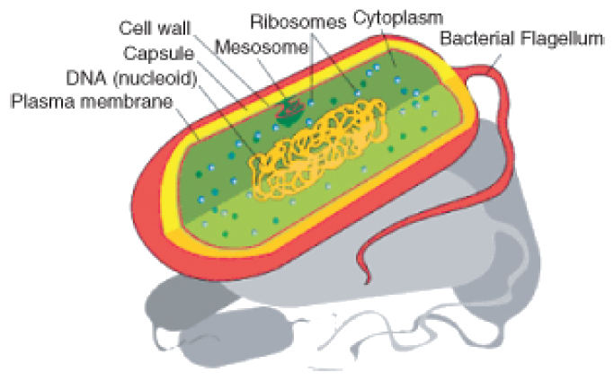
Figure 8: Diagram of a typical prokaryotic cell. Among other things, prokaryotic cells have a plasma membrane, cytoplasm, ribosomes, and DNA. Prokaryotes do not have membrane-bound organelles or a cell nucleus.
Most prokaryotes are unicellular and have a cell wall that adds structural support and acts as a barrier against outside forces. Some prokaryotes have an extra layer outside their cell wall called a capsule, which helps them stick to surfaces or to each other. Prokaryotic DNA usually forms a circular molecule and is found in the cell’s cytoplasm along with ribosomes. Prokaryotic cells are very small; most are between 1–10 μm in diameter. They are found living in almost every environment on Earth. Biologists believe that prokaryotes were the first type of cells on Earth and that they are the most common organisms on Earth today.
Eukaryotic Cells
A eukaryote is an organism whose cells are organized into complex structures by internal membranes and a cytoskeleton, as shown in Figure 9. The most characteristic membrane-bound structure of eukaryotes is the nucleus. This feature gives them their name, which comes from Greek and means ”true nucleus.” The nucleus is the membrane-enclosed organelle that contains DNA. Eukaryotic DNA is organized in one or more linear molecules, called chromosomes. Some eukaryotes are single-celled, but many are multicellular.
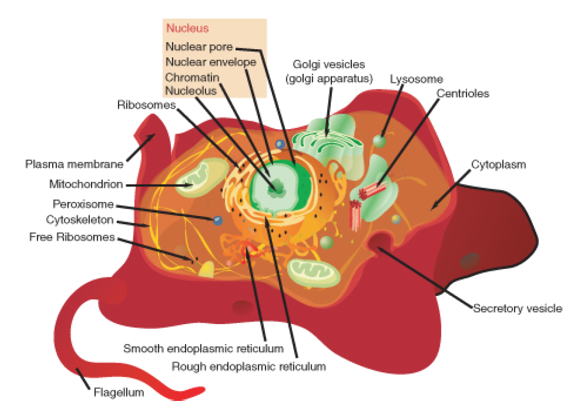
Figure 9: A eukaryotic cell, represented here by a model animal cell is much more complex than a prokaryotic cell. Eukaryotic cells contain many organelles that do specific jobs. No single eukaryotic cell has all the organelles shown here, and this model shows all eukaryotic organelles.
In addition to having a plasma membrane, cytoplasm, a nucleus and ribosomes, eukaryotic cells also contain membrane-bound organelles. Each organelle in a eukaryote has a distinct function. Because of their complex level of organization, eukaryotic cells can carry out many more functions than prokaryotic cells. The main differences between prokaryotic and eukaryotic cells are shown in Figure 11 and listed in Table 1. Eukaryotic cells may or may not have a cell wall. Plant cells generally have cell walls, while animal cells do not.
Eukaryotic cells are about 10 times the size of a typical prokaryote; they range between 10 and 100 μm in diameter while prokaryotes range between 1 and 10 μm in diameter, as shown in Figure 10. Scientists believe that eukaryotes developed about 1.6 – 2.1 billion years ago. The earliest fossils of multicellular organisms that have been found are 1.2 billion years

Figure 10: The relative scale of prokaryotic and eukaryotic cells. See how eukaryotic cells are generally 10 to 100 times larger than prokaryotic cells.

Figure 11: The main differences between prokaryotic and eukaryotic cells. Eukaryotic cells have membrane bound organelles while prokaryotic cells do not.
Table 1 Structural Differences Between Prokaryotic Cells and Eukaryotic Cells
| Presence of | Prokaryote | Eukaryote |
|---|---|---|
| Plasma Membrane | yes | yes |
| Genetic Material (DNA) | yes | yes |
| Cytoplasm | yes | yes |
| Ribosomes | yes | yes |
| Nucleus | no | yes |
| Nucleolus | no | yes |
| Mitochondria | no | yes |
| Other membrane-bound organells | no | yes |
| Cell wall | yes | some (animals cells do not have it) |
| Capsule | yes | no |
| $$\text{Average diameter}$$ | 0.4 to 10 μm | 1 to 100 μm |
Are Viruses Prokaryotic or Eukaryotic?
Are viruses prokaryotic or eukaryotic? Neither. Viruses are not made up of cells, so they do not have a cell membrane or any cytoplasm, ribosomes, or other organelles. Viruses do not replicate by themselves, instead, they use their host cell to make more of themselves. So most virologists consider viruses non-living. But, they do evolve, which is a characteristic of living things.
A virus is a sub-microscopic particle that can infect living cells. Viruses are much smaller than prokaryotic organisms. In essence, a virus is simply a nucleic acid surrounded by a protein coat.

Figure 12: Structural overview of a virus, the T2 phage. A 2-dimensional representation is on the left, and a 3-dimensional representation is on the right. The virus is essentially nucleic acid surrounded by a protein coat.
Images courtesy of:
Robert Hooke, Micrographia,1665. Suber cells and mimosa leaves. Public Domain.
http://en.wikipedia.org/wiki/Image:Antoni_van_Leeuwenhoek.png. Public Domain.
Dr.Ralf Wagner. http://commons.wikimedia.org/wiki/Image:Rotifera.jpg. GFDL.
http://commons.wikimedia.org/wiki/File:Elektronenmikroskop.jpg. (a)Public Domain (b)GNU-FDL.
Niamh Gray-Wilson. CC-BY-SA.
Raul654,JWSchmidt. http://en.wikipedia.org/wiki/Image:Ostrich_egg.jpg. GNU-FDL,GNU-FDL.
http://commons.wikimedia.org/wiki/Image:Redbloodcells.jpg
http://remf.dartmouth.edu/images/bacteriaSEM/source/1.html
http://remf.dartmouth.edu/images/algaeSEM/source/1.html
http://remf.dartmouth.edu/images/botanicalPollenSEM/source/10.html.
CC-BY,Public Domain,Public Domain,Public Domain,Public Domain.
http://en.wikipedia.org/wiki/Image:Prokaryote_cell_diagram.svg. Public Domain.
http://en.wikipedia.org/wiki/Image:Animal_cell_structure.svg. Public Domain.
http://en.wikipedia.org/wiki/Image:Relative_scale.png. Public Domain.
CK-12 Foundation. http://schools-wikipedia.org/images/915/91506.png.htm. Public Domain.
http://en.wikipedia.org/wiki/Image:Tevenphage.png. CC-BY-SA 2.5.